S-WAVE Software
The Software Behind Every Scan
From the scan at home to your dentist’s review: how the S-WAVE™ software is being designed.
Concept only. The S-WAVE™ software does not exist yet. The screens below are design concepts with sample data; features, phases and data commitments describe intent and may change. S-WAVE™ is in development, has not been tested in people and is not cleared or approved by the FDA.
A look inside
One Workspace, Every Step
Scan, explore in 3D, review AI findings and plan treatment without switching programs.
S-WAVE · Sample patient (demo data) · Full-arch scan · 3 Nov 2026
 3D viewer
 Live scan
 AI findings
 Perio chart
 Implant planner
 Compare
 Biomarkers
 Share & reports
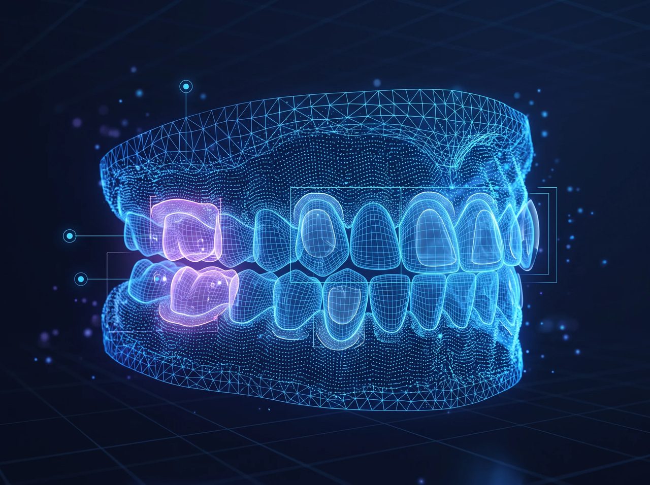
AI findings
3 to review- 1#3 · #14Review
Bone level below normal
Distal crest 3.1 mm from CEJ
Confidence87% - 2#24Review
Gingival recession
1.4 mm on the buccal side
Confidence92% - 3#30Review
Implant site ready to plan
Bone width 6.8 mm · height 11.2 mm
Confidence95%
 Suggestions only — the clinician confirms every finding.
Illustration with sample data. Features are shown by development phase and may change.
Workflow
From Scan to Shared Plan
- 01

Capture
One full-arch ultrasound scan with the tray.
- 02

Reconstruct
AI turns the echoes into a 3D model in seconds.
- 03

Analyse
Findings flagged for the clinician to review.
- 04

Plan
Measure, chart and plan implants on the model.
- 05

Share
Export STL/DICOM, send to specialists and patients.
Capture
Phase 1Live Scan Guidance
The software guides every scan as it happens, so the first scan is a usable scan — no retakes, no guesswork for the assistant.
 Live coverage map that fills in tooth by tooth
- Signal quality and tray seating checks in real time
- Clear prompts when an area needs a moment longer
Live scan · Upper arch
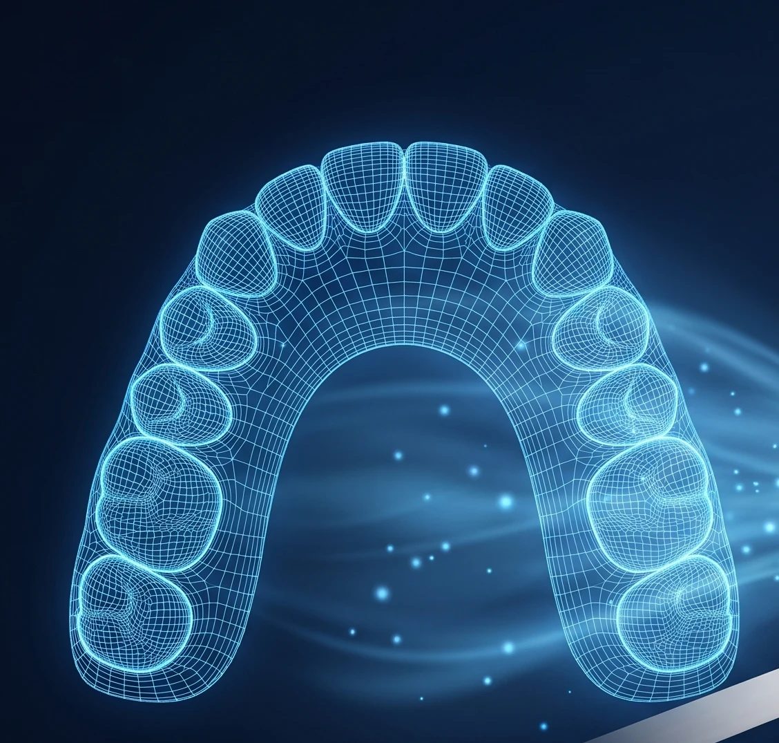
Signal quality
Excellent
Tray seating
Correct
Teeth captured
Hold still 3 more seconds for #13–#15.
Explore
Phase 13D Viewer with Tissue Layers
Ultrasound sees below the surface. Turn layers on and off, slice through any tooth and measure in tenths of a millimetre.
- Enamel, roots, gums, bone and implants as separate layers
- Cross-sections through any tooth, any angle
- Distance, angle and thickness measurements
3D viewer · Cross-section through #19
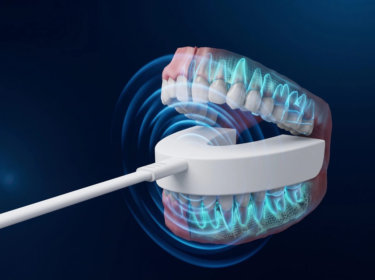
Tissue layers
Diagnose
Phase 2AI Findings, Clinician in Control
AI looks at every scan the same careful way and points out what deserves a second look. Each finding shows where, how much and how sure — the dentist decides.
- Bone level, gum recession and soft-tissue changes
- Confidence score and exact location for each finding
- Accept, dismiss or edit — every decision is logged
AI findings
- 1#3 · #14Review
Bone level below normal
Distal crest 3.1 mm from CEJ
Confidence87% - 2#24Review
Gingival recession
1.4 mm on the buccal side
Confidence92% - 3#30Review
Implant site ready to plan
Bone width 6.8 mm · height 11.2 mm
Confidence95%
Chart
Phase 2Periodontal Charting in Seconds
Pocket depths and gum levels are measured from the scan and filled into a standard perio chart — no probing round, no dictation.
- Six sites per tooth, buccal and lingual
- Problem sites highlighted automatically
- Edit any value by hand before saving
Periodontal chart · auto-filled from scan
| Buccal (D · B · M) | Lingual (D · L · M) | ||||||
|---|---|---|---|---|---|---|---|
| #2 | 3 | 4 | 3 | 2 | 3 | 3 | |
| #3 | 5 | 6 | 4 | 3 | 4 | 4 | |
| #4 | 3 | 2 | 3 | 2 | 2 | 3 | |
| #5 | 2 | 3 | 2 | 2 | 2 | 2 | |
| #6 | 3 | 3 | 2 | 2 | 3 | 2 | |
| #7 | 2 | 2 | 3 | 3 | 2 | 2 | |
2.9
mean depth
2
sites ≥5 mm
4 s
to chart
Plan
Phase 2Implant Planning
Bone width, height and soft tissue are measured where the implant will go, with the distance to the nerve always in view.
- Bone and soft-tissue measurements at the site
- Implant size suggestion with safety margins
- Plan export for surgical guides
Implant planner · Site #30
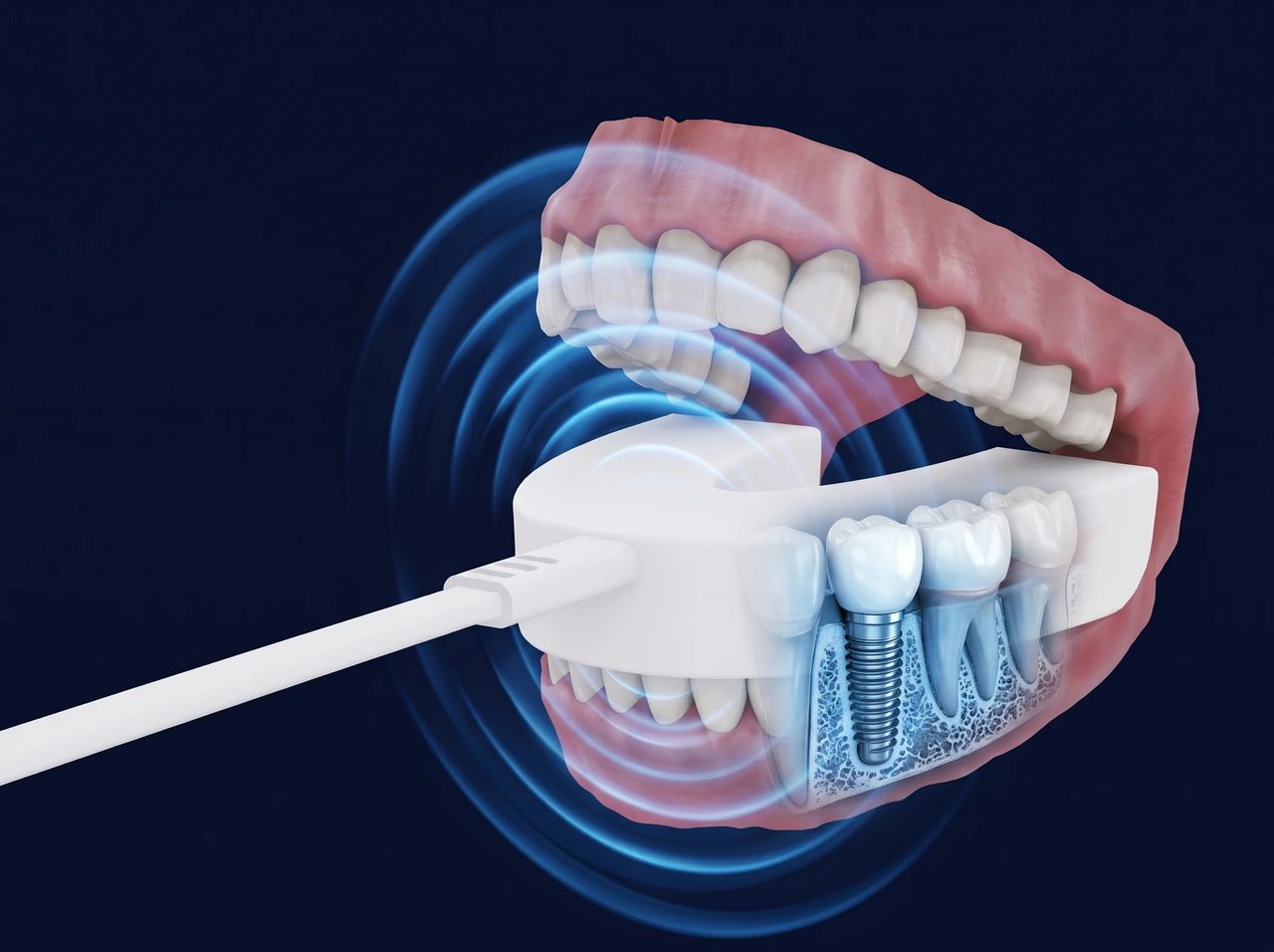
Suggested implant
Ø 4.1 × 10 mm
Safety margin to nerve
2.4 mm ✓
Soft tissue thickness
2.9 mm
Monitor
Phase 2Compare Scans Over Time
Radiation-free means you can scan at every visit. The software lines up two scans and shows exactly what changed.
- Automatic alignment of scans from different visits
- Change map in millimetres
- Progress charts patients understand
Compare scans · May 2026 → Nov 2026
| Measure | May | Nov | Change |
|---|---|---|---|
| Gingival recession #24 | 0.6 mm | 1.4 mm | +0.8 mm |
| Pocket depth #3 (M) | 5 mm | 4 mm | −1 mm |
| Bone level #14 (D) | 2.8 mm | 3.1 mm | +0.3 mm |
| Plaque index | 38% | 12% | −26% |
Screen
Phase 3Saliva & Breath Biomarkers
A lab-on-chip cartridge reads saliva and breath markers chairside and adds them to the patient’s record next to the scan.
- pH, inflammation markers, glucose and breath VSCs
- Results in minutes, trends across visits
- Early warning for gum disease and systemic risk
Biomarkers · lab-on-chip test · 4 minutes
Saliva pH
Normal6.8
normal 6.5–7.5
Inflammation marker
WatchElevated
IL-1β trend ↑
Breath VSC
Normal142 ppb
normal < 150
Glucose (saliva)
Normal0.9 mg/dL
reference range
Collaborate
Phase 2Tele-dentistry & Reports
Send a case to a specialist or the lab in one click, and give patients a clear report they can open on their phone.
- Secure, expiring share links with comments
- Plain-language patient reports
- Remote second opinions without moving the patient
Share case
Periodontist · referral
3D model, perio chart, 2 findings
Dental lab
STL upper + lower, bite
Patient report
Plain-language summary, phone friendly
Encrypted link · expires in 14 daysSendOpen by design
Exports & Integrations
Your data is never locked in. Every scan leaves the software in open, standard formats.
STL · PLY · OBJ
3D models for any CAD/CAM software, 3D printer or milling machine.
DICOM
For CBCT software and fusion with existing X-ray volumes.
PDF reports
Clinical and patient reports with your practice details.
Practice software
Link scans and findings to the patient record (planned).
Privacy & security
Built for Patient Data
Health data deserves the strictest care. These are the principles the platform is built on.

Encrypted
Scans encrypted in transfer and at rest.

Role-based access
Dentist, assistant, lab and patient each see only what they need.

Audit trail
Every view, edit and share is recorded.
Works offline
Scan and review at the chair; sync when connected.
Everywhere you work
One Platform, Four Screens
Chairside tablet
Runs the scan with the tray, live guidance and instant 3D.
Desktop workstation
Full diagnosis, charting and planning on a big screen.
Web portal
Specialists and labs open shared cases in the browser.
Patient view
Reports and progress on the patient’s phone.
Roadmap
Where the Software Is Going
Features arrive in phases, starting with scanning and 3D, then AI diagnosis and planning, then biomarkers and real-time sensors.
Phase 1
Scan & see
- Full-arch ultrasound capture
- Live scan guidance
- 3D viewer and tissue layers
- STL / DICOM export
Phase 2
Diagnose & plan
- AI findings
- Automatic perio charting
- Implant planning
- Scan comparison
- Sharing and reports
Phase 3
Monitor & predict
- Saliva and breath biomarkers
- CBCT fusion and elastography
- Chairside real-time sensors
- Risk prediction across visits
Dental imaging without radiation, from home.
Questions about S-WAVE™, the research or a collaboration? Our team is here to help.