Technology
A full-arch 3D ultrasound scan, at home, in minutes
How the tray works, what the bench prototype already does, and the software and regulatory work ahead.
How it works
Photos stop at the surface. Ultrasound goes below it.
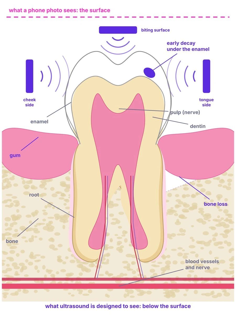
A phone photo shows
- Tooth position and alignment
- Stains and visible plaque
- Nothing under the enamel or the gum line
S-WAVE™ is designed to show
- Early decay under the surface
- Gum thickness and bone level around every root
- Enamel, dentin and pulp, from three sides
- No X-rays, no radiation
One scan a month. A dentist is alerted only when something changes.
Months 1–6: nothing to do. Month 7: a change below the gum line is flagged; the dentist calls the patient in while the fix is still small.
Three steps, at home
Minutes at home. All sensors fire at once: the whole arch in one shot, no probe sweep, no X-rays.
AI being designed to flag change early, before it becomes an emergency.
Telehealth dentists contracted by KronosMD, or your own practice.
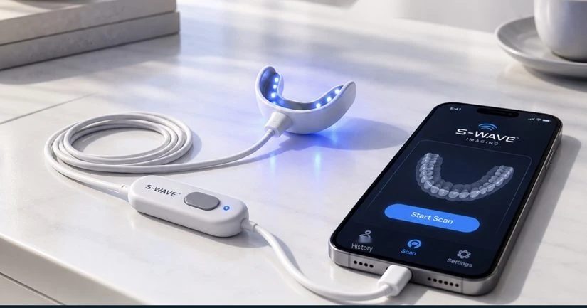
A tray lined with ultrasound sensors and a camera, wired to your phone.
Illustration, not a clinical image. Capabilities are design objectives supported by published studies of research-grade dental ultrasound (see Science); S-WAVE™ itself has not been tested in people. S-WAVE™ supports monitoring between visits and does not replace examination, diagnosis or treatment by a licensed dentist. Planned use, subject to FDA authorization.
Inside the scan
What S-WAVE™ Is Designed to See
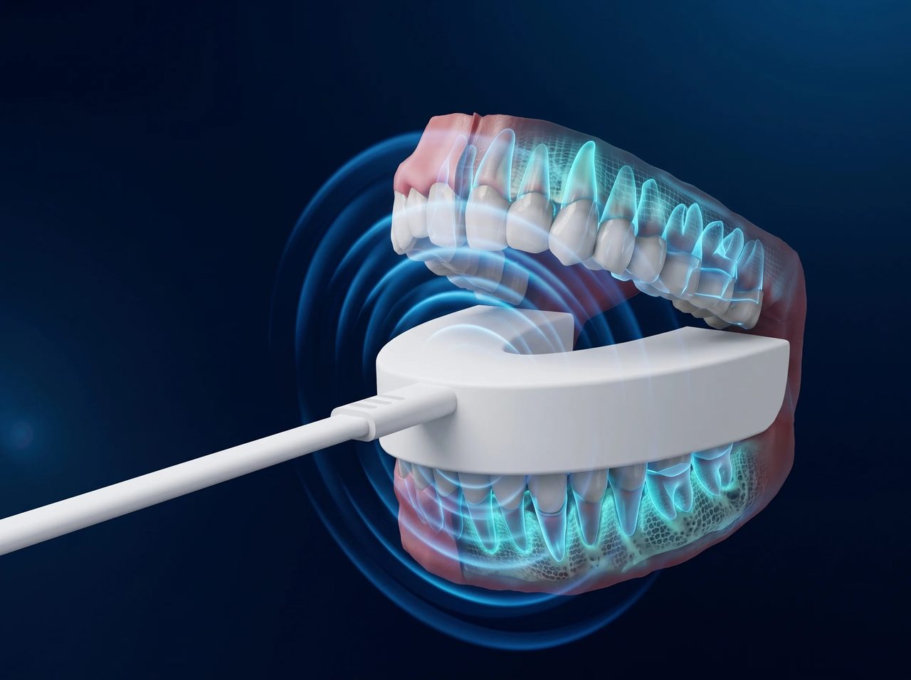
Ultrasound doesn’t stop at the surface. S-WAVE™ is designed to image teeth, gums, bone and implants together, and turn the echoes into a 3D model.
Through the gums
Ultrasound passes through gum tissue to show roots, the periodontal ligament and bone — what optical scanners can’t see.
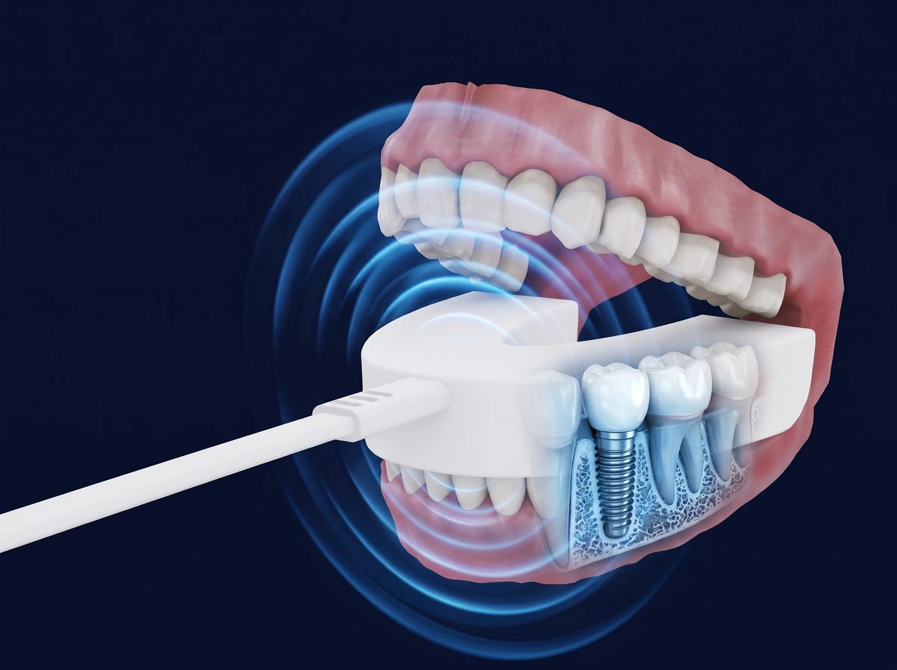
Around implants & metal
Designed to read past crowns and metal restorations, which block X-rays. Implant imaging is a future research direction.
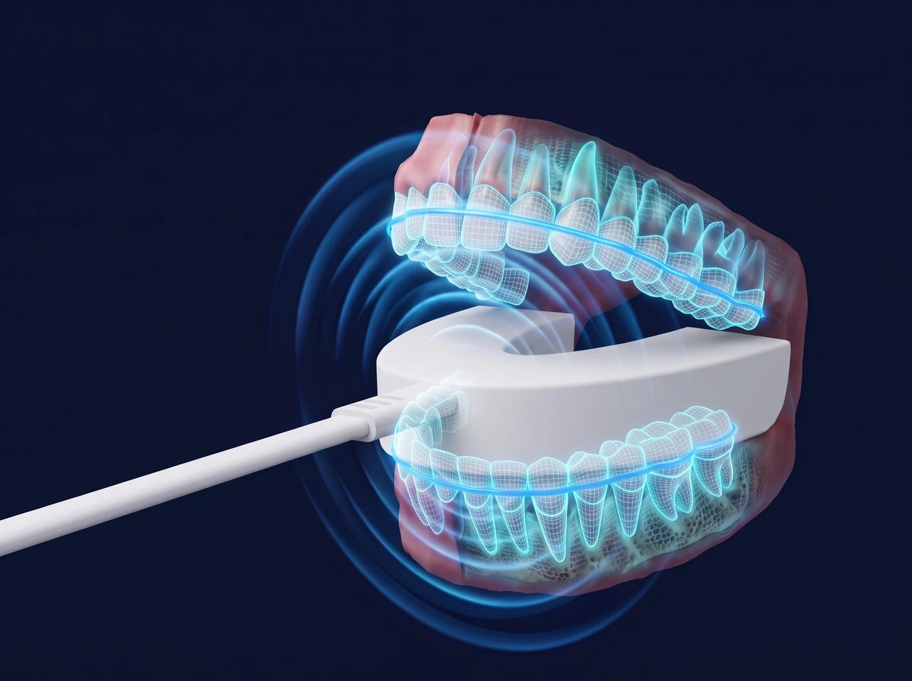
The full arch at once
The tray is designed to capture the whole arch in one scan: no wand, no repeated passes.
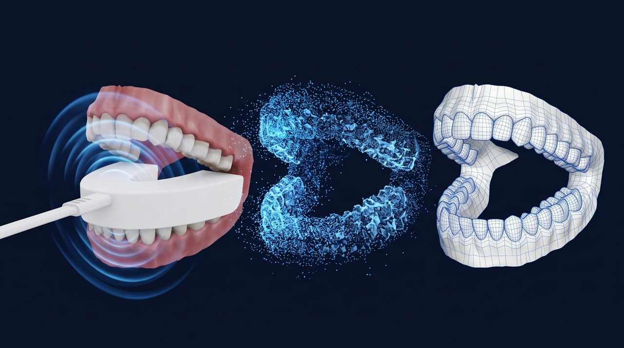
From echo to 3D model
AI Reconstruction
Deep learning turns the ultrasound echoes into a cloud of points and then a precise 3D model of the arch, exported as STL and DICOM for your CAD/CAM workflow.
Illustrations, not clinical images. Capabilities are design objectives; S-WAVE™ has not been tested in people.
The principle
From one probe to a full arch: why a tray, not a wand
Every published dental ultrasound study used one probe
A clinician sweeps a single transducer over one surface of one tooth and the system stitches 2D slices. Results depend on the operator: in one enamel study, one examiner reached r = 0.97 and another r = 0.47.
S-WAVE™ fixes the geometry
A tray lines the cheek side, the tongue side and the biting surface of every tooth with sensors. They fire together, so the whole arch is captured from multiple angles in a single acquisition, with no sweep, no stitching and no operator skill required.
Borrowed from industry and medicine
Full-matrix capture and total-focusing reconstruction inspect aircraft and nuclear components; ultrasound tomography rings the breast with transducers to build a true 3D volume (FDA-approved 2021); NASA rates pulse-echo ultrasound “quantitative with size and depth information.” S-WAVE™ is designed to apply the same idea to the mouth.
From raw echoes to a monthly comparison: the software we plan to build
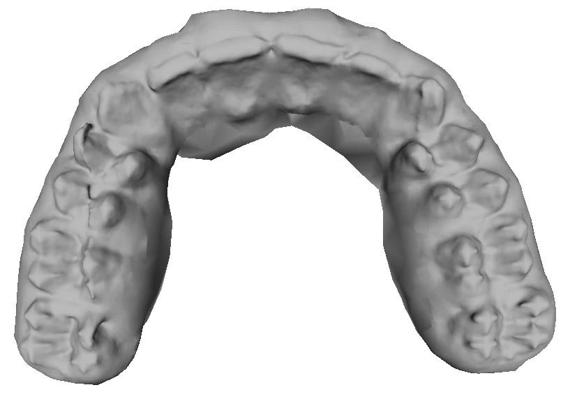
Echoes from every sensor position become a point cloud, a mesh and a surface. The bench prototype does this today; the raw result is shown here.
AI being designed to remove noise, separate teeth from gum and bone, and label each tooth.
Each scan is aligned to last month’s and differences are measured: enamel, gum thickness, bone level.
Only changes above a threshold are sent for review. The dentist, not the software, decides what to do.
Industrial and medical systems named here are not KronosMD products and their results do not predict S-WAVE™’s. Multi-angle 3D reconstruction of a full arch in a single acquisition is a design objective that has not been demonstrated in people. Sources: TWI; Eddyfi; FDA PMA P200040; NASA/TM-2020-220568; NASA Spinoff.
Proof
It already works on the bench: a full arch in 3D
Real data
A full upper arch rendered straight from ultrasound echoes: no X-rays, no optical scanner, and no AI processing yet. Cusps, fissures and the curve of the palate are already visible.
How it was made
Produced in the laboratory by the 3D bench scanner prototype acquired with the IP and technology. Echoes from multiple sensor positions were reconstructed into a point cloud, a mesh and this surface.
What comes next
The planned AI is designed to process raw scans like this one: clean and segment the surface, register each scan to the last, and flag change for a dentist. Not the consumer tray, not a patient, not a clinical result.
A foundational patent
US 10,799,210: cited by 11 later filings
An intraoral scan head combining an ultrasonic transducer with a light-based image sensor, producing 3D dental images. Later patent applications by Colgate-Palmolive, Carestream/Trophy, Konica Minolta and the University of California cite it as prior art. Origin: US 6,589,054 (filed 2000, now expired), in which an Oregon dentist and a lumber engineer adapted stress-wave defect detection from timber inspection to teeth. New provisional filings are planned after the raise.
Where the technology came from
A separate company, an acquired technology
KronosMD acquired the patents, prototype and research data in 2022 from the shareholders of the technology’s original developer, paying in KronosMD shares. KronosMD is a separate company with no affiliation to that developer. Details and risk factors are in the Form C.
Raw image produced in the laboratory by the 3D bench scanner prototype, not by the consumer device, which has not been built. Not a patient; not a clinical result; not tested in people. Patent citations per USPTO forward-citation records as reviewed by the Company; a citation does not imply licensing, infringement or endorsement.
Design targets
What we are engineering toward
| Conventional impressions | In-office intraoral scanner | S-WAVE™ (design target) | |
|---|---|---|---|
| Where | Dental office | Dental office | Home |
| Who operates | Staff | Trained staff | The patient; fixed tray, no sweep |
| Time | ~67 min | ~25 min | Minutes (target) |
| Sub-surface imaging | No | No | Yes (design goal) |
| Ionizing radiation | None | None | None |
| Connection | — | Cart or USB | Wired to the patient’s phone; no radio in the tray |
| Sensors | — | Optical | Thin-film ultrasound on buccal, lingual and occlusal walls, plus a camera for color |
| Tested in people | Standard of care | FDA-cleared products | Not yet |
Conventional-impression and scanner figures are Company estimates and have not been independently verified. S-WAVE™ values are design targets, not measured results.
Regulatory plan and roadmap
Home-use authorization comes first
Ask FDA to confirm the pathway (510(k) Class II vs De Novo) for an intraoral ultrasound imaging device intended for home use with remote clinician review.
Acoustic output and safety, biocompatibility, software verification and validation, AI performance and clinical accuracy versus reference imaging, lay-user human factors.
eSTAR submission with a predetermined change control plan for the AI software. Imaging and monitoring indication only.
Consumer-direct and practice channels; ongoing software updates under the change control plan.
Where we are today
Images a full arch in 3D on the bench. Electronics being updated for demonstration and miniaturization.
Bench and scan data acquired with the IP: the basis for AI development and a planned peer-reviewed paper.
US 10,799,210, active, cited by 11 later filings. New provisional filings planned after the raise.
Dr. Greg Rubin. Engineering and software hires on closing at least $1,000,000; product design partner engaged under a written agreement.
What we have not done
No testing in people. No FDA clearance. No commercial agreements. No revenue. Very little cash: the Company depends on this offering to keep operating. We would rather you heard that from us first.
Beyond imaging: a platform that can add services over time
Sensors in the tray designed to read saliva biomarkers during a scan. Research stage.
Acoustic energy intended to activate whitening agents at home under remote supervision. Research stage.
Focused ultrasound intended to support delivery of periodontal medicaments. Research stage.
Ultrasound-assisted remineralization intended to arrest very early cavities. Research stage.
Target dates are estimates and may be delayed; the FDA could require more testing than we expect, or may never authorize S-WAVE™; consumer home use may face a higher bar. Platform add-ons are not part of the current product or this offering, which funds the imaging and monitoring product only; a therapeutic or diagnostic add-on would be a different regulatory product requiring clinical trials and its own FDA authorization, and the Company has conducted no such trials.
Dental imaging without radiation, from home.
Questions about S-WAVE™, the research or a collaboration? Our team is here to help.